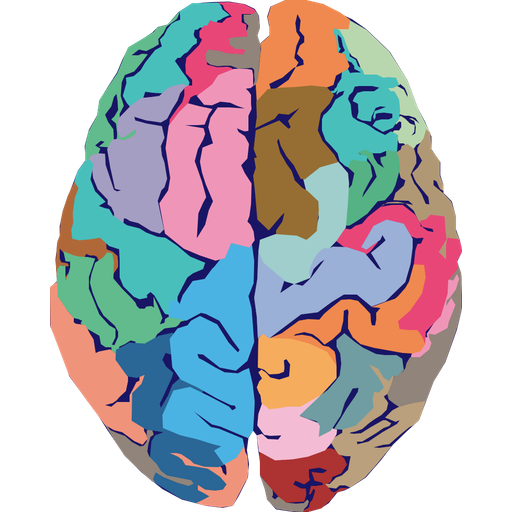
# cover page, Hae-Jeong Park, Jae Jin Kim, Seung-Koo Lee, Jeong Ho Seok, Jiwon Chun, Dong Ik Kim and Jong Doo Lee, Corpus Callosal Connection Mapping Using Cortical Gray Matter Parcellation and DT-MRI, Human Brain Mapp, 29, 503–516, 2008.
[here]표지 논문
한글 요약
좌우 대뇌반구를 연결하는 뇌량의 어느 부위가 어느 대뇌피질 영역과 이어지는지를 사람 집단 수준에서 지도화한 연구입니다. 건강한 성인의 구조 MRI에서 대뇌피질을 수십 개 영역으로 자동 분할하고, 이를 확산텐서영상(DT-MRI) 섬유 추적과 결합하여 각 피질 영역에서 출발한 뇌량 섬유가 정중 시상면의 어느 지점을 통과하는지를 확률 지도로 만들었습니다. 그 결과 전전두엽 섬유는 뇌량 앞부분인 무릎과 부리를, 두정엽과 후두엽 섬유는 뒷부분인 팽대를, 감각운동피질 섬유는 협부를 주로 지나며, 대체로 등쪽·안쪽 피질은 뇌량의 등쪽을, 가쪽 피질은 배쪽을 통과하는 규칙성이 확인되었습니다. 이렇게 피질 연결에 근거해 뇌량을 세분하면 기존의 기하학적 구분보다 더 정밀하게 뇌량 구조를 이해할 수 있습니다.
초록 (English)
Population maps of the corpus callosum (CC) and cortical lobe connections were generated by combining cortical gray matter parcellation with the diffusion tensor fiber tractography of individual subjects. This method is based on the fact that the cortical lobes of both hemispheres are interconnected by the corpus callosal fibers. T1-weighted structural MRIs and diffusion tensor MRIs (DT-MRI) of 22 right-handed, healthy subjects were used. Forty-seven cortical parcellations in the dorsal prefrontal cortex, ventral prefrontal cortex, sensory-motor cortex, parietal cortex, temporal cortex, and occipital cortex were semi-automatically derived from structural MRIs, registered to DT-MRI, and used to identify callosal fibers. The probabilistic connections to each cortex were mapped on entire mid-sagittal CC voxels that had anatomical homology between subjects as determined by spatial registration. According to the population maps of the callosal connections, the ventral prefrontal cortex and parts of the dorsal prefrontal cortex both project fibers through the genu and rostrum. The CC regions through which the superior frontal cortex passes extend into the posterior body. Fibers arising from the parietal lobe and occipital lobe run mainly through the splenium, while fibers arising from the sensory-motor cortex pass through the isthmus. In general, dorsal or medial cortical lobes project fibers through the dorsal region of the CC, while lateral cortical lobes project fibers through the ventral region of the CC. The probabilistic subdivision of the CC by connecting cortical gray matter provides a more precise understanding of the CC.